

July '26
A new cellular compartment: the atonosome
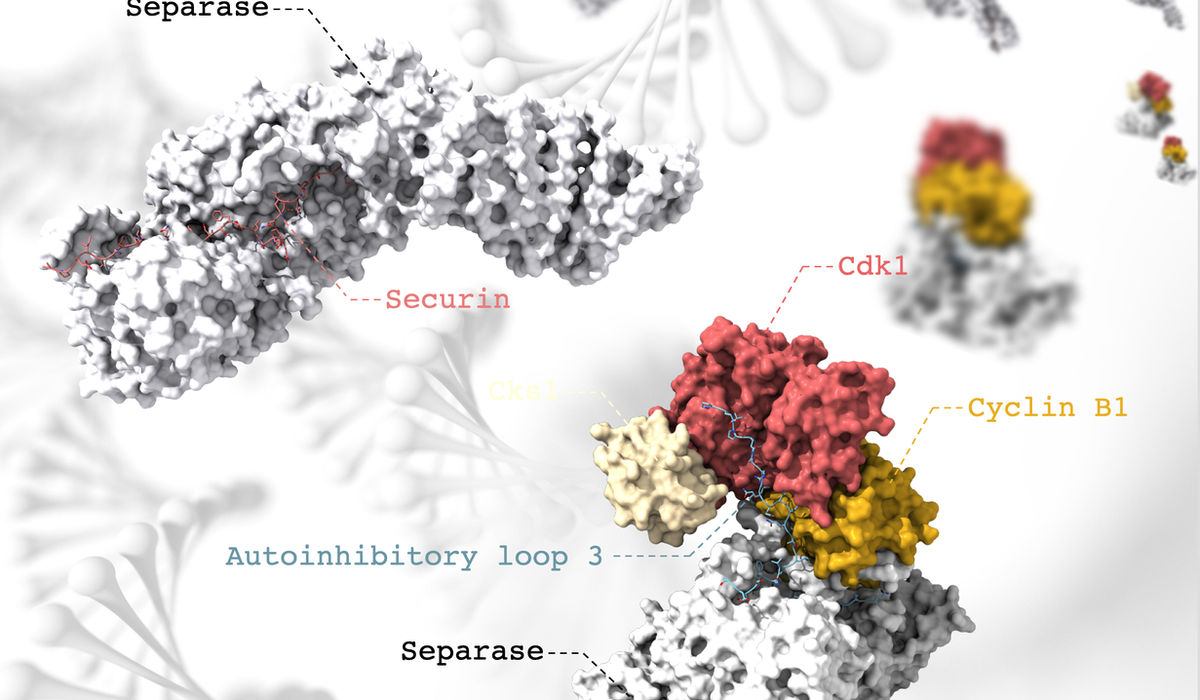
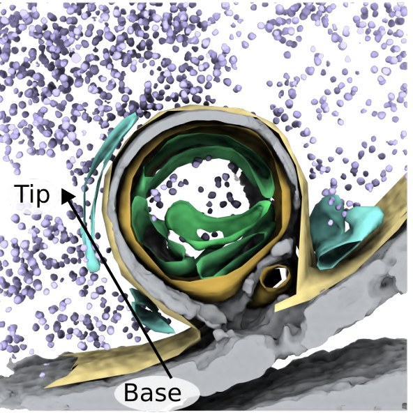
Elda Bauda's recent work uncovered a new cellular compartment we call the "atonosome," which forms within seconds when a cell's outer membrane loses tension — such as during osmotic shock. Using high-resolution cryo-ET of yeast, we show these structures arise across contexts of both acute and chronic membrane-tension loss. Posted as a preprint on bioRxiv; not yet peer-reviewed.
Read the preprint →